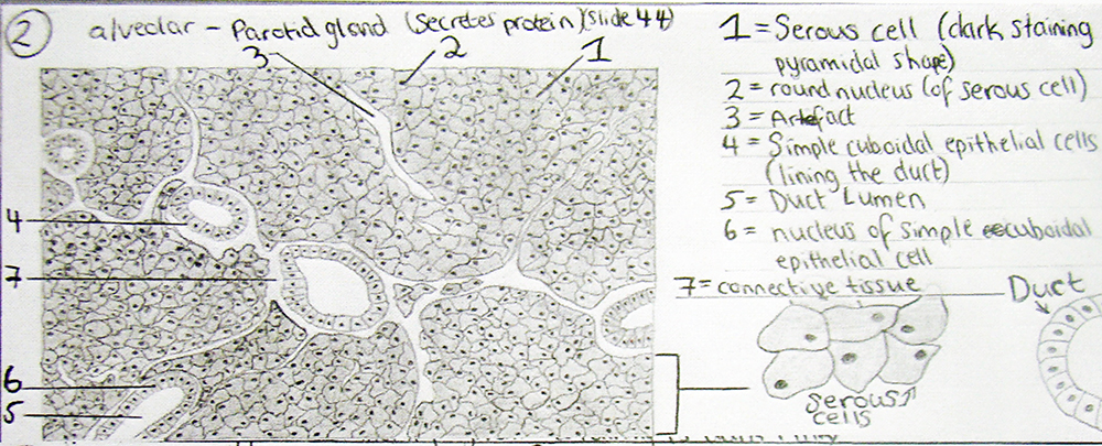
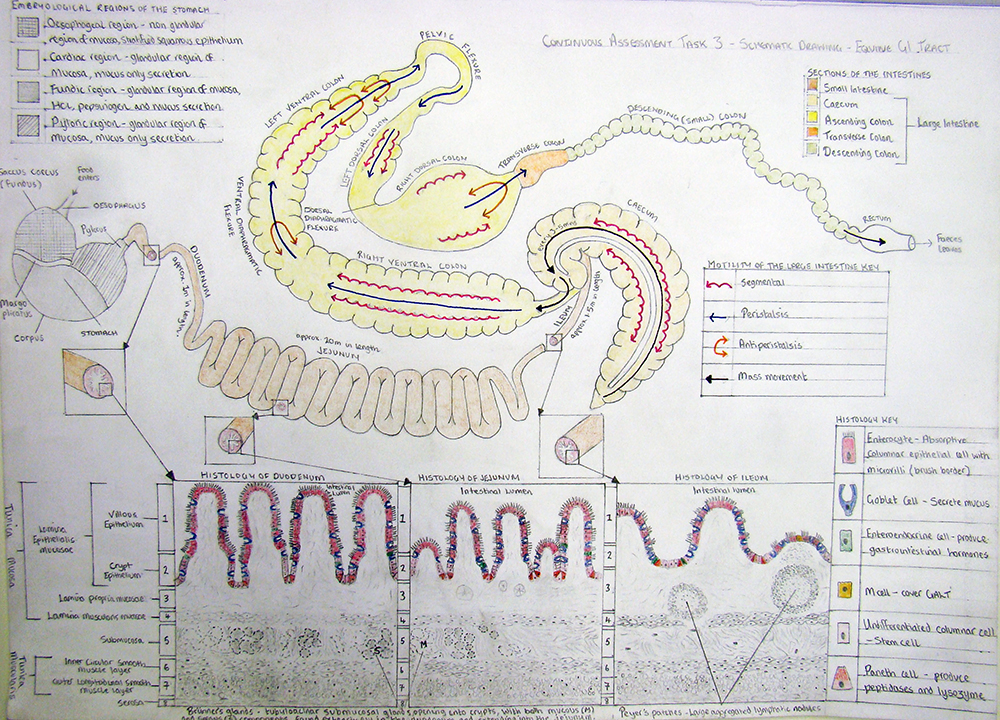
Using Drawing as a Way of Understanding: University of Liverpool Veterinary Science Schematic Drawing Task.
Many thanks to Fay Penrose, Lecturer in Veterinary Biology at The Veterinary School at University of Liverpool, who shares her work in introducing drawing as a way of checking understanding of complex subjects. This methodology would be very transferable to a number of subject areas in schools.

Introduction
Anatomy, the science that concerns the form of the tissues and organs of the body, is by its very nature a visual and tactile science. A good understanding of the normal shape and colour of things, their texture and how they relate to other structures in space (topographically) aids understanding of normal function and the ability to recognise abnormal, or pathological, processes. Medical, dental and veterinary undergraduate students spend a large part of their first two years at university learning anatomy to enable them to become good clinicians.
However, anatomy is not easy to learn. There is so much of it and everything has a name. Or several different names. Even things that don’t exist, potential spaces, have names. Every organ or structure has specific parameters of normal shape and colour, which students become familiar with by looking and touching specimens during practical classes. Away from these classes students need to find a way to summarise their understanding in order to clarify, revise and explain functional anatomical concepts.
Aims
Traditionally many students learn or revise by creating written notes which are time consuming to produce and often unclear or contain contradictory or vague statements. Revisiting them for the purposes of revision is also time consuming and requires the reader to ‘translate’ the written word into a mental visual image. Acknowledged as a problem by veterinary staff and students alike is the sheer volume of material that has to be learnt. In the context of clinical anatomy, merely memorizing the names or shapes of things is not enough, an understanding of their functional importance is vital to become a problem solving practitioner. This short series of tasks asked students to draw instead of write. We asked them to use observational and conceptual skills to represent an example each of microanatomy, skeletal anatomy and to summarise their understanding of an anatomical system. The aim was to condense information given to students during several lectures and practical classes that had been delivered over the course of one semester. The overarching objective was to encourage students to use drawing as an efficient and clear way to communicate their knowledge of form and function.
The Task
Three separate tasks were set as compulsory exercises that formed part of the continuous assessment strand for first year veterinary science undergraduates. Students were asked to submit a histology drawing, an anatomical drawing and a schematic drawing. The first two tasks involved drawing from observation, whilst the third task summarised concepts. All three tasks were limited to one side of A4 paper. The histology (micro-anatomy) task focused on depicting the cell structure of different types of gland and required observation down a microscope. In addition, this task was limited to grayscale colours to allow the students to focus on the microstructure and spatial relationships. The anatomical drawing task, also in grayscale, involved drawing directly from skeletal specimens in the laboratory. Finally, with the schematic task we asked students to convey the complexity of the equine gastrointestinal tract. For this task as well as overall shape and size, they had to convey the embryological derivation of the stomach, the histology of the small intestine and the motility of the large intestine. As this final drawing was schematic it could be portrayed as ‘life-like’ shapes or simply as straight lines and boxes. However, whichever method the individual student chose the length and proportions of the different parts of the tract had to be accurate. In essence all three tasks asked students to convey a lot of detailed and specialised information in a small space. Students were instructed to use minimal text, and consider use of colour or shading/hatching/stippling to differentiate different regions or tissues.


Lessons Learned
When the details of the tasks were released a large number of students expressed the opinion that they could not draw. However, the results demonstrated otherwise – all students can draw! Some students commented that they felt they had been pushed outside of their comfort zone but actually appreciated doing the exercises and would consider summarising their learning in a similar fashion in future. Although the students needed to invest a reasonable amount of time to produce accurate drawings they were learning whilst they did so and by the end, had produced valuable resources that they could use as an ‘at a glance’ revision guide. A few students also commented that they could see how it was a useful skill to cultivate – not only for their own understanding but also to communicate with future patients/clients.


